Whenever a sample from an immunocompromised patient comes around, there's a good chance we'd find something uncommon. Lo and behold, my colleague found something under the scope.
He suspected that it could be Cryptococcus neoformans. The suspicion alone was enough for us to take different precautions with the patient's culture. Trying to relive the old school days, he wanted to see if we had any India ink to visualize the potential fungus under the microscope. Funnily enough, we procured some ink from the histology department.
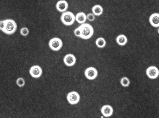
The pictures I tried to take were horrendous. They were the equivalent of taking shots of the moon with your smartphone. The white dots were supposed to be the fungus.
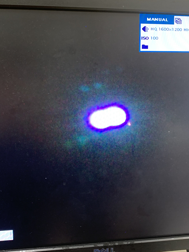
As expected, bringing the image to an external display didn't work either. The contrast alone made visualization impossible without witnessing it yourself.
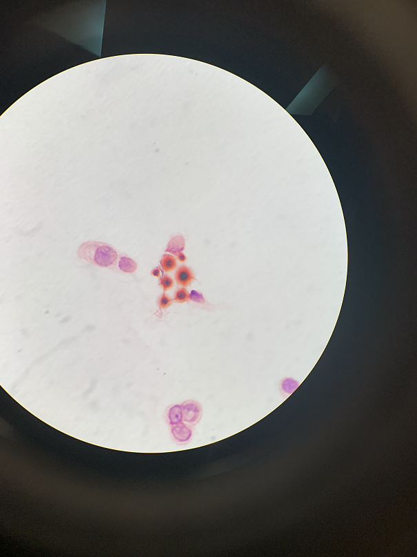
source
The picture above is what you are supposed to see under the scope. I hope you can make sense of the terrible photos from earlier with this representation.
The last thing we needed to do was set up fungal cultures for this patient. In addition, we moved the routine cultures off of automation. They had to be sealed with parafilm and could only open under BS2 environment. In days' time, we can confirm the identity of the fungus we saw in the stains.
Cryptococcus neoforman is an interesting species. It's a radiotrophic fungus that can thrive in extreme radioactive conditions. I recall there was little information online about radiosynthesis 10 years ago.
Fortunately for us, this particular fungus is usually an opportunistic infection. I still wouldn't go breathing them in, though. You can read more about it on the CDC website.
Posted with STEMGeeks